
The Experimental Immunology Lab
Current Team: Verena Labi (PI), Katia Schöler (PhD candidate), Katharina Hoppe (PhD candidate), Sarah Spöck (PhD candidate), Johannes Wölk (MSc candidate), Ilaria Dorigatti (MSc candidate), Nadine Kinz (MSc candidate)

Pathologies of the immune system represent a major threat to healthy aging because of their abundance and limited therapeutic options.
In our group, we study molecular mechanisms that control cell survival and fate decisions in B lymphocytes and their progenitors, through genetic mouse models and in vitro culture systems. Since 2015 we have identified the integration of miRNAs in B cell survival and selection, with the miR-17-92 miRNAs playing key roles in promoting autoimmunity. A similar principle holds in the control of malignant B cell outgrowth and MYC-driven lymphoma cell fitness where the miR-17-92 target gene BIM takes center stage. Separate lines of work have revealed CHK1-mediated DNA damage control as key element of normal B cell development, germinal center B cell fitness and antibody-mediated immunity. Recent achievements include the identification and characterization of the epigenetic TET enzymes as critical regulators of B-cell lineage specification and optimal humoral immune responses. Additional new insights relate to the tumor suppressor functions of TET2 operating in hematopoietic progenitors and B lymphocytes.
Graphical abstract of our discoveries on the immune system in normal physiology and therapy (involved molecules are indicated in red/bold).

MAJOR ACHIEVEMENTS AND ONGOING RESEARCH EFFORTS INCLUDE:
APOPTOSIS AS BARRIER TO AUTOIMMUNITY AND CANCER
The identification of the pro-apoptotic Bcl2-protein Bmf as specific regulator of B cell fitness and mediator of therapy-induced cell death (HDACi, glucocorticoids). Building on these results we discovered that Bmf and its relative Bim synergize in promoting programmed apoptosis in the developing embryo and in the adult immune system – the latter function efficiently preventing autoimmunity and cancer, both in and beyond the immune system.
FEBS J, doi: 10.1111/febs.14426; JEM, doi: 10.1084/jem.20071658; Oncogene, doi: 10.1038/onc.2009.42; Blood, doi: 10.1182/blood-2013-11-537217; Cell Death Differ, doi: 10.1038/cdd.2015.8; Blood, doi: 10.1182/blood-2009-03-212670; Cell Death Differ, PMID: 16645634
APOPTOSIS IMPAIRS GRAFT QUALITY AND TRANSPLANTATION OUTCOME
The finding that Bim and Bmf are rate-limiting for homeostatic hematopoietic stem and progenitor cell survival in mice and men with broader implications for transplantation therapy in humans. Ongoing research efforts focus at establishing procedures to enhance graft quality ex vivo.
EMBO Mol Med, doi: 10.1002/emmm.201201235; JEM, doi:10.1084/jem.20161721
DNA DAMAGE SIGNALING IN B CELLS
The discovery that developing and antigen-activated B cells exert control over the DNA damage response while introducing mutations in their antibody genes via fine-tuning Chk1 concentration. Our results predict that therapeutic Chk1 inhibition in cancer patients may prove potent in killing B cell lymphoma and leukemia cells addicted to B cell receptor signaling.
Nat. comm., doi: 10.1038/s41467-017-01850-4; Cell Death Differ, doi: 10.1038/s41418-019-0318-5
EPIGENETIC REGULATION OF B CELL IDENTITY AND FUNCTION
The first description of active DNA demethylation as a requirement for tissue development in the adult. Combined lack of TET2 and TET3 from early B cell development on prevents focal DNA hypomethylation at enhancers and consequently corrupts the gene expression program associated with maturation transitions. Consequently, the resulting mature B cells are non-functional. Current studies seek to clarify whether TET-function controls antibody-mediated immunity as well as the mutational spectrum in germinal center B cells and B cell lymphomas.
PNAS, doi: 10.1073/pnas.1604365113; FEBS J, doi: 10.1111/febs.14934
A miRNA CLUSTER CONTROLS B CELL FITNESS AND PATHOLOGY
The identification of a PI3K-driven c-Myc/miR-17~92/Pten autostimulatory loop that critically controls selection of B cell progenitors into the mature B cell pool. Recently, we identified the Rag-genes, essential for developmental DNA recombination in early B cell progenitors, as targets of this regulatory pathway. In an ongoing project, we are dissecting the vital and diverse impact of the miR-17~92 cluster through control of a single apoptotic target gene in normal and malignant B cells and beyond the immune system.
G&D, doi: 10.1101/gad.330134; Cell Reports, doi: 10.1016/j.celrep.2016.05.084; Front Immunol, doi: 10.3389/fimmu.2018.02715; Cancer Lett, doi: 10.1016/j.canlet.2018.12.020
A PARADOX: APOPTOSIS ENFORCING TUMORIGENESIS
Classically, the ability of cells to undergo apoptosis has been established as a major barrier against cancer. Contrasting common beliefs, one of our most exciting findings is that apoptosis can promote cancer under certain circumstances. A model emerged where waves of apoptosis in peripheral lymphatic organs foster transformation of damaged hematopoietic stem and progenitor cells by enforcing proliferation to compensate for the loss of cells in the periphery (see Figure 2 below). This finding has been recapitulated by others in different cancer models, (e.g. HCC, MDS-AML transition), observations that underline the importance of our inaugural findings for human cancer pathology, but also with significant implications for therapy-induced malignancies. Future research aims to understand the molecular and physiological causes and consequences of cell death for homeostatic turnover, tissue injury-repair, pre-neoplastic transformation and cancer progression.
G&D, doi: 10.1101/gad; Cell Cycle, PMID: 20980816; Cell Death Dis, doi: 10.1038/cddis.2015.20
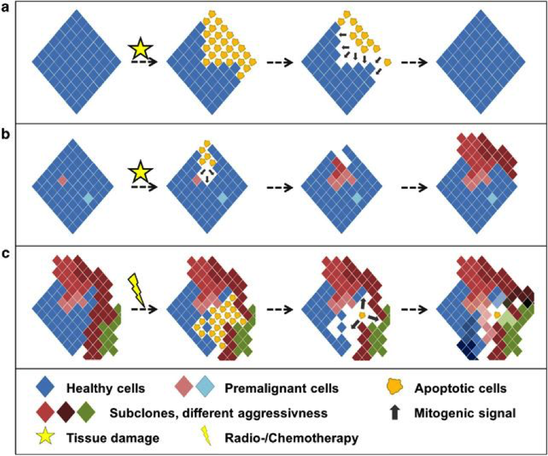
Figure 2: How apoptosis shapes tissues and cancer.
(a) In proliferative tissues, injury is followed by rapid regeneration and restoration of normally sized and shaped structures. In the Drosophila wing imaginal disc, apoptotic cells induce competitive proliferation by secretion of mitogenic factors in a caspase-dependent manner. (b) In tissues with aberrant cells, tissue injury (e.g. caused by DNA damage in MDS patients) and consecutive proliferation enables outgrowth of more aggressive clones. This fosters malignant transformation. (c) Within established tumors, chemo- or radiotherapy induces apoptosis but leads to death-induced proliferation of therapy-surviving cells. Following the generation of space, proliferation is mediated by mitogens derived from apoptotic cells (such as PGE2). As proposed in mathematical models, this results in increased sub-clonal variability with a higher risk of tumor progression, chemoresistance and relapse. Cell Death Dis, doi: 10.1038/cddis.2015.20